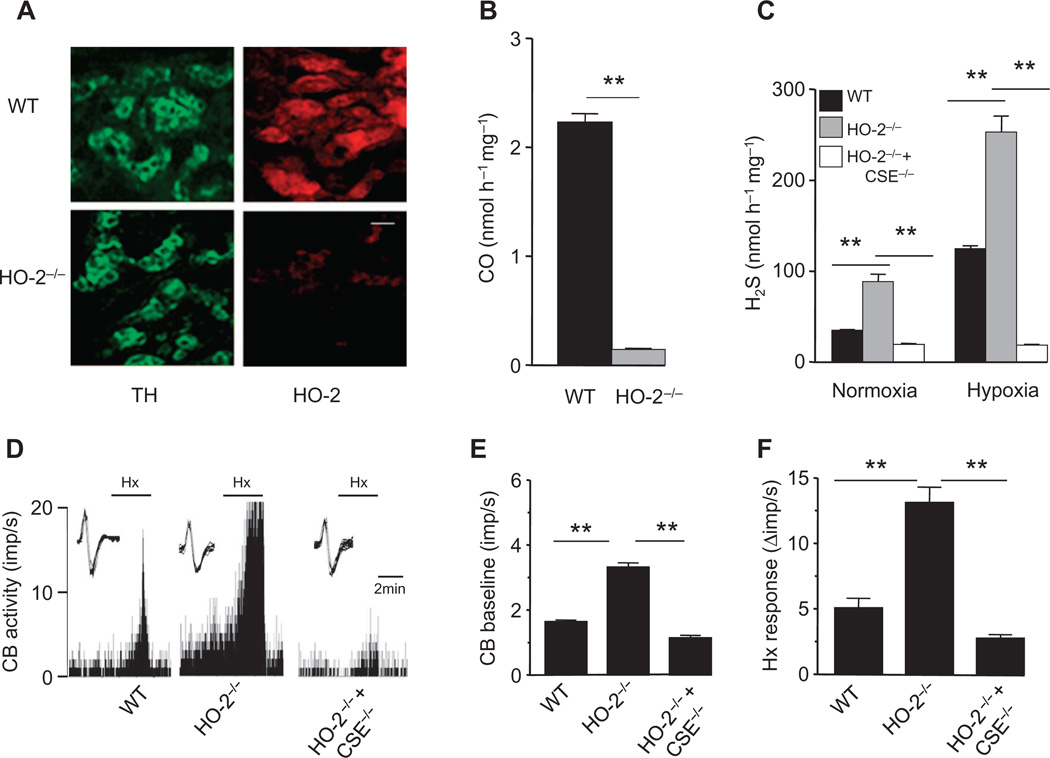
Fig. 5. Gaseous messenger generation and sensory nerve activity in HO-2–null carotid bodies.
(A) Adjacent carotid body sections immunostained for HO-2 and tyrosine hydroxylase (TH), a marker of glomus cells, in WT and HO-2−/− mice. Scale bar, 20 µm. (B) CO generation in carotid bodies of WT and HO-2−/− mice. (C to F) H2S generation (C) and sensory nerve activity (D to F) of the carotid bodies of WT, HO-2−/−, and HO-2−/− + CSE−/− mice. In tracings of (D), the insets present superimposed action potentials of the sensory nerve fiber from which the integrated carotid body sensory nerve activity (CB activity; imp/s) was derived, and black bars marked with “Hx” represent the duration of the hypoxic challenge. Images in (A) are representative of three mice per genotype. The graphs in (B), (C), (E), and (F) represent means ± SEM (n = 3 independent experiments for CO and H2S measurements and n = 6 carotid bodies for each genotype for sensory nerve activity measurements). **P < 0.01.

