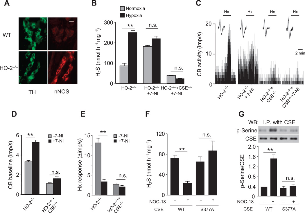
Fig. 6. NO signaling in HO-2–null carotid body.
(A) Adjacent carotid body sections immunostained for nNOS and tyrosine hydroxylase (TH), amarker of glomus cells, inWT and HO-2−/− mice. Scale bar, 20 µm. (B to E) Effect of the nNOS inhibitor 7-NI on H2S generation (B) and sensory nerve activity (C to E) of carotid bodies from HO-2−/− and HO-2−/− + CSE−/− mice. In tracings in (C), the insets present superimposed action potentials of the sensory nerve fiber from which the integrated carotid body sensory nerve activity (CB activity; imp/s) was derived, and black bars marked with “Hx” represent the duration of the hypoxic challenge. (F and G) Effect of the NO donor NOC-18 on H2S generation (F) and CSE serine phosphorylation (G) in HEK-293 cells expressing WT or mutant CSE (S377A). Images in (A) are representative of three mice per genotype. The graphs in (B) and (D) to (G) represent means ± SEM (n = 3 for each genotype and treatment for H2S measurements, n = 6 to 8 carotid bodies for each genotype and treatment for sensory nerve activity measurements, and n = 3 independent experiments for H2S measurements and CSE phosphorylation in HEK-293 cells). **P < 0.01.

