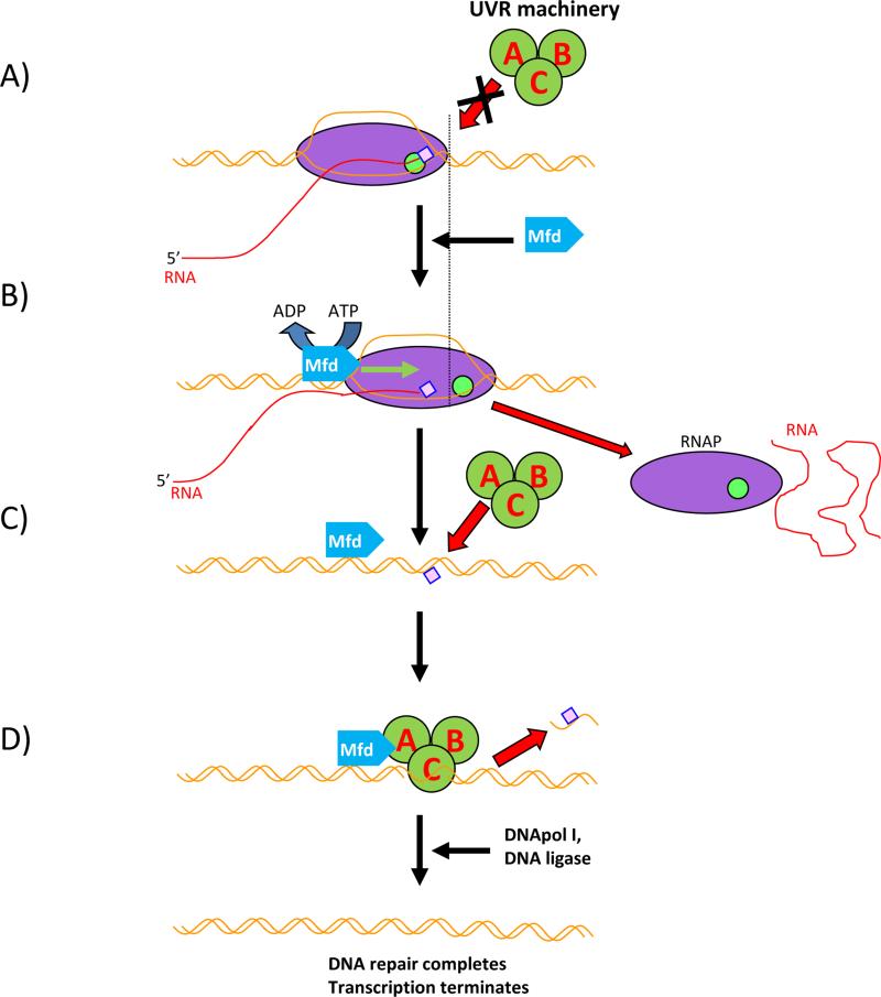
Figure 1.
Scheme of Mfd-depended TCR in bacteria. RNAP – magenta oval, active site – green circle, Mfd – blue pentagon. UvrABC system – dark green circles, bulky lesion - pink square. DNA – brown line, RNA – red line. A: RNAP encounters a bulky lesion on the template strand and stalls, unable to elongate. Elongation Complex blocks UvrABC from loading onto the DNA. B: Mfd recognises stalled Elongation Complex, loads upstream and pushes RNAP forward by using its helicase/translocase activity. Eventually RNAP is removed, RNA synthesis is terminated and the lesion becomes exposed. C: The UvrABC system is actively recruited at the DNA damage site by Mfd-UvrA interactions and UvrABCD machinery removes the lesion. D: UvrABCD leave the complex; DNApol and DNA ligase finish DNA repair.

