
Figure 1.
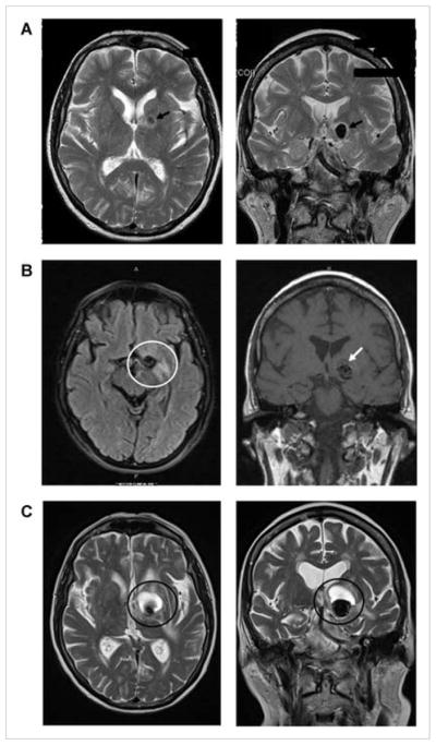
Axial (left column) and coronal (right column) T2 weighted (A, C) and fluid attenuated inversion recovery (FLAIR) MRI images (B) demonstrating aneurysm evolution: (A) prior to intervention, a 12-mm left internal carotid artery terminus aneurysm (black arrows) anterior and superior to the left cerebral peduncle; (B) 14 months after endovascular coil embolization with parenchymal T2 signal hyperintensity around the left callosal and pericallosal regions of the coiled aneurysm site (white circle) and some low signal at the aneurysm apex (white arrow), above the coil mass (represented as area of signal loss) on the coronal view. Low signal is inconsistent with blood product and likely represents developing cyst (T2 images not obtained); (C) 15 months after repeat endovascular coil embolization with enlarged aneurysm and superior expansion of a cystic mass compressing internal capsule, pallidum, and some caudate (dark circles).