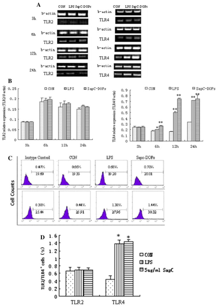
Fig. 4.
Effects of SapC-DOPS on TLR2 and TLR4 expression. Raw264.7 cells were treated with PBS or 5 μg/ml SapC-DOPS, LPS (0.5 μg/ml) and PBS as positive control. a After incubation with SapC-DOPS or LPS for 3, 6, 12 or 24 h, Raw264.7 cells were collected and TLR2 or TLR4 mRNA were determined by RT-PCR. b Densitometric analysis. The intensity of the band was scanned. The quotients of TLR2 and TLR4/β-actin gene were calculated. The results are shown as mean ± SE from three representative independent experiments. c, d The TLR2 or TLR4 protein level in macrophages after incubating with 5 μg/ml SapC-DOPS or 0.5 μg/ml LPS were measured with flow cytometry analysis. Results were expressed as means ± SD from three independent experiments. *p < 0.05, **p <0.01 versus control

