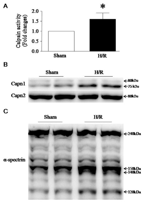
Fig. 4.
Measurement of calpain activation in H9c2 cells following H/R. H9c2 cells were subjected to a 24-hour hypoxia followed by a 24-hour re-oxygenation. (A) Calpain enzymatic activity was measured in H9c2 cells. Data are mean ± SD from 3 different experiments. *P < 0.05. (B) is a representative western blot for a 75 kDa active fragment of calpain-1 and (C) for the cleavage of α-spectrin (140 kDa), a natural substrate of calpain-1.

