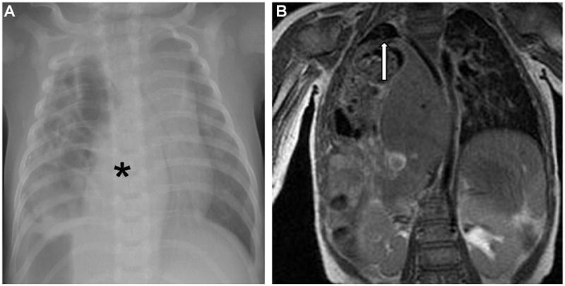
Figure 1.

Figure 1A – Chest radiograph at time of presentation demonstrated a right sided diaphragmatic hernia with bowel in right chest and contralateral mediastinal shift. Black asterisk denotes liver in thorax.
Figure 1B – Coronal magnetic resonance image of patient’s chest. In right chest, liver is seen herniating and area of fusion to underdeveloped right lung is denoted by white arrow.
