Figure 2.

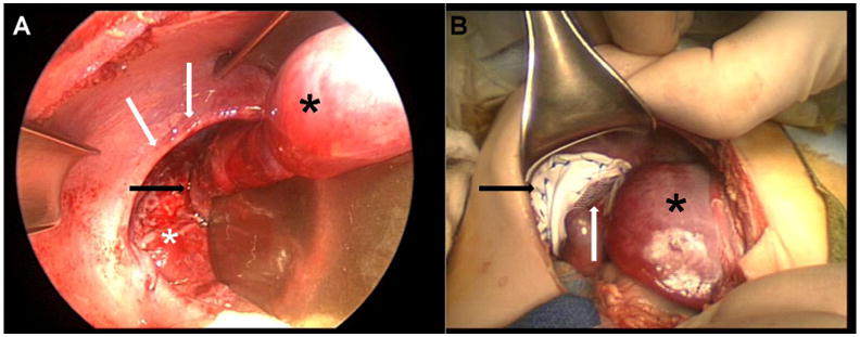
Figure 2A – View from abdomen. Black arrow denotes site of hepatic pulmonary fusion. White arrows show border of diaphragmatic defect. White asterisk on lung and black asterisk on liver. Retractor is seen at right side of image.
Figure 2B – After approximation of diaphragmatic defect. Black asterisk is liver, black arrow is PTFE mesh and white arrow is polyester fiber mesh connecting liver to the PTFE patch.
