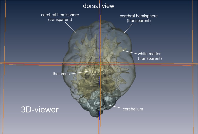
Fig 3. Volume rendering of brain tissues of interest.
3D viewer mode of the graphical software AMIRA. The voxels of the tissue of interest (white matter/grey matter) of each slice have been assembled and are now displayed as a 3D model. Each tissue can be displayed solid or transparent. The localizer lines support the segmentation process. As they are displayed in both the 2D images and the 3D model, the thalamus, medulla and cerebellum can be accurately separated from the volume of interest.

