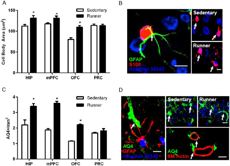
Fig 2. Running alters astrocyte morphology in regions associated with increased cognitive performance.
A, S100+ astrocyte cell body area is increased in the hippocampus, medial prefrontal cortex, and orbitofrontal cortex. B, Left: S100+ astrocyte (red) colabeled with GFAP (green). Scale bar = 5 μm. Right: Representative images of astrocytes from the medial prefrontal cortex of sedentary and running animals. Scale bar = 10 μm. C, Optical density of aquaporin-4, a water channel found in the endfeet of astrocytes, was increased in the hippocampus, medial prefrontal cortex, and orbitofrontal cortex of runners. D, Left: Aquaporin-4 (green) colabels with GFAP (red). Scale bar = 5 μm. Right top: Representative images of aquaporin-4 expression in CA1 of sedentary and running animals. Scale Bar = 20 μm. Right bottom: aquaporin-4 labeling (green) is shown in close proximity to smooth muscle actin labeling (red). Scale bar = 20 μm. Error bars represent SEM. *p<0.05 compared with Sedentary for A and C.

