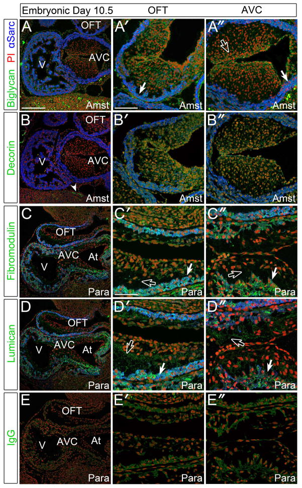
Figure 1. Biglycan, fibromodulin, and lumican were detected in the myocardium and endocardial cushions of E10.5 murine embryonic hearts.
A–A″: Immunolocalization of biglycan (green) in Amsterdam (Amst) fixed tissue. B–B″: Decorin (green) immunolocalization in Amst fixed tissue. C–C″: Fibromodulin (green) staining in paraformaldehyde (Para) fixed tissue. D–D″: Immunolocalization of lumican (green) in Para fixative. E–E″: IgG (green) controls in sister sections. Solid arrows- myocardial staining; open arrows- endocardial cushion staining; arrowhead- non-cardiac DCN immunoreactivity. V- ventricle; AVC- atrioventricular cushions; OFT- outflow tract; At- atrium. Blue- alpha sarcomeric actin; red- propidium iodide. Scale bars: A= 200um, applies to B, C, D, E; A′= 100um, applies to A″, B′, B″; C′= 50um, applies to C″, D′, D″, E′, E″.

