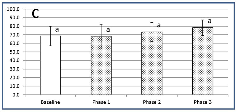
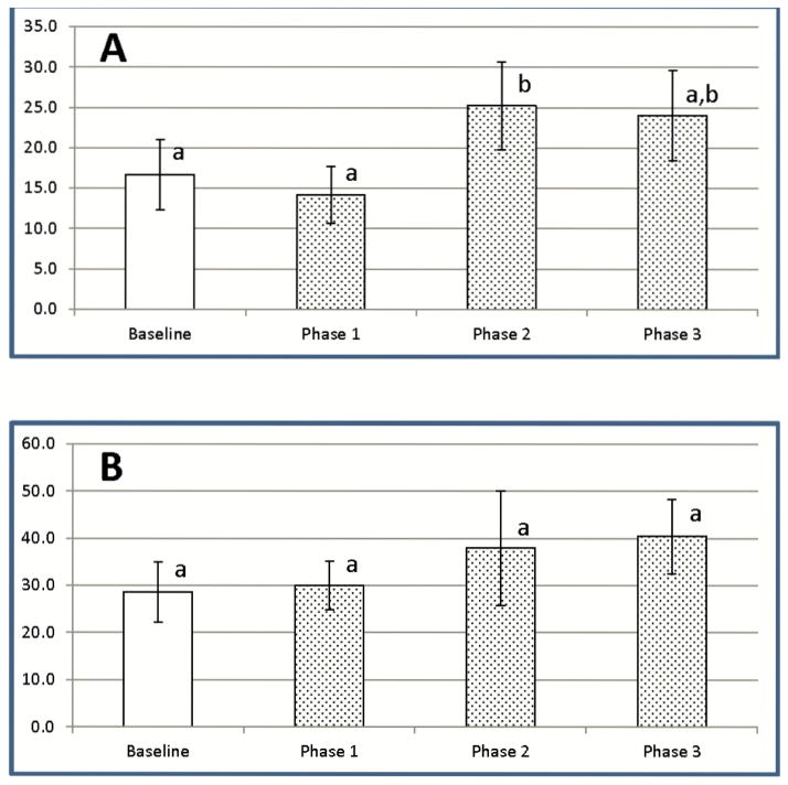
Figure 7.

LC exposure promotes significant increases in the most recently activated CD4+CD25lowFOXP3+ Tregs. CD4+FOXP3+ T cells from the dogs exposed to LC were gated based on brightness of CD25 staining resulting in separation into low, medium, and high expressers of CD25. (A) – Low CD25 expressors (B) – Medium CD25 expressors (C) – High CD25 expressors. Data are presented as % FOXP3+Tregs of CD4+T cells with low, medium and high CD25+ expression (mean ± SD). a,b Means containing the same superscript are not significantly different from each other (P<0.05).

