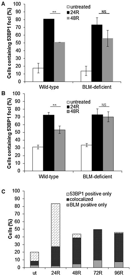
Fig. 4.
Accumulation and co-localization of 53BP1 and BLM foci following formaldehyde treatment. Kinetics of 53BP1 (A) and H2AX (B) foci accumulation in wild-type and BLM-deficient cells following formaldehyde treatment. Cells were treated with formaldehyde (300 μM for 4 h), fixed following a 24 and 48 h recovery (24R and 48R, respectively), and stained with anti-53BP1 or γH2AX antibody. The mean and standard deviation from three or more independent experiments are shown. Significant difference (Student’s t-test, **P ≤ 0.01); NS, not significant. (C) Co-localization of 53BP1 and BLM in wild-type cells following formaldehyde exposure. Under the same treatment conditions as used for 53BP1 staining, cells were co-stained with anti-BLM and anti-53BP1 antibodies to follow the co-localization of the two proteins over a 4 day recovery period (R). For each sample, 100 cells were counted in every experiment. The mean of three or more independent experiments are shown. “ut” denotes untreated cells.

