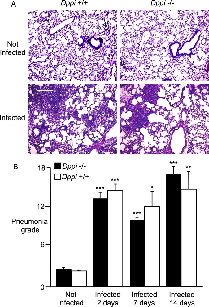
Fig 2. Comparison of mycoplasma tracheobronchitis and pneumonia in Dppi-/- and Dppi+/+ mice.
A: photomicrographs of hematoxylin- and eosin-stained lung sections from wild type (Dppi +/+) and Dppi-deficient (Dppi -/-) control mice (“Not Infected”) or mice infected 2 days previously with Mycoplasma pulmonis. Scale bar = 240 μm. B: comparisons of quantitative grading of airway and parenchymal inflammation (“Pneumonia grade”) in control mice (“Not Infected”) and mycoplasma-infected mice whose lungs were harvested 2, 7 and 14 days after infection; *P = 0.05, **P = 0.01, and ***P < 0.001 in comparison to “Not Infected” controls; N = 4–7 mice per group.

