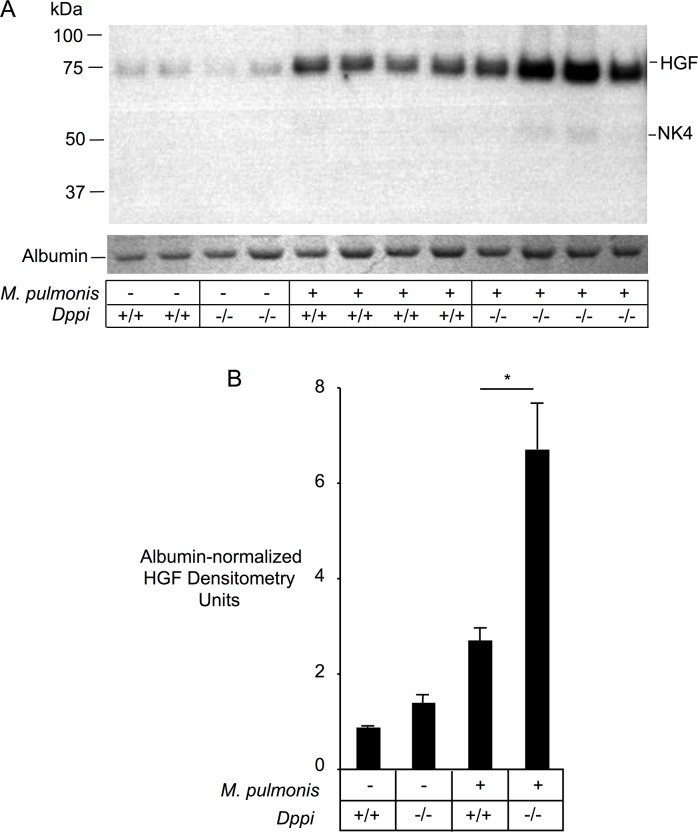
Fig 4. HGF and NK4-like fragments in Mycoplasma pulmonis-infected Dppi-/- and Dppi+/+ mice.
Panel A shows anti-HGF immunoblots of BALF proteins in parallel with albumin bands from a Coomassie blue-stained SDS-PAGE gel of the same samples. The panel also shows immunoblots of HGF in serum collected 2 and 7 days after mycoplasma infection. Panel B shows results of densitometry of HGF bands normalized to albumin band intensity. Infected mice were lavaged 24 hours after receiving a nasal inoculum of 0.5x10⁶ M. pulmonis.

