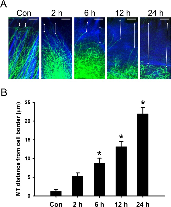
Fig 1. HYS-32 prevents microtubules from targeting to cell cortex.
(A) Control astrocytes (Con) or astrocytes treated for 2, 6, 12, or 24 h with 5 μM HYS-32 were fixed in cold acetone and double-stained for β -tubulin (green) and F-actin (blue). Double arrows indicate the distance between microtubule tips and the cell border (bars = 5 μm). (B) Quantitative analyses of the distance between microtubule tips and cell border were performed as described in Materials and Methods. The results were collected from three independent experiments. *p<0.01compared to control (Con) using one-way ANOVA with Dunnett’s post-hoctest.

