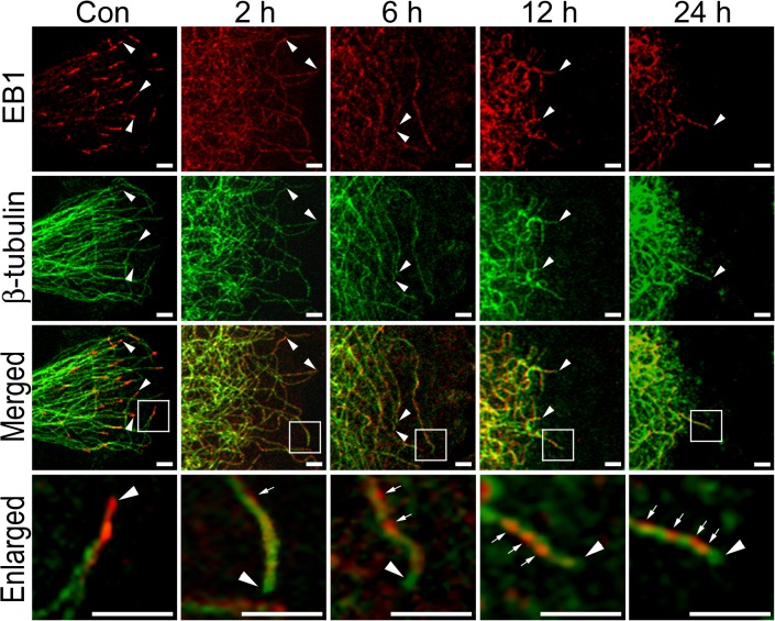
Fig 2. HYS-32 induces extensive distribution of EB1 on the microtubule lattice and eliminates EB1 staining from the microtubule plus end.
Control astrocytes (Con) or astrocytes treated for 2, 6, 12, or 24 h with 5 μM HYS-32 were fixed in cold acetone and double-stained for β-tubulin (green) and EB1 (red) and subjected to confocal microscopy. Images were merged to show co-localization (Merged). Square areas were enlarged to show EB1 distribution (Enlarged). Arrowheads indicate microtubule tips. Arrows indicate distribution of EB1 along the microtubules (bars = 5 μm).

