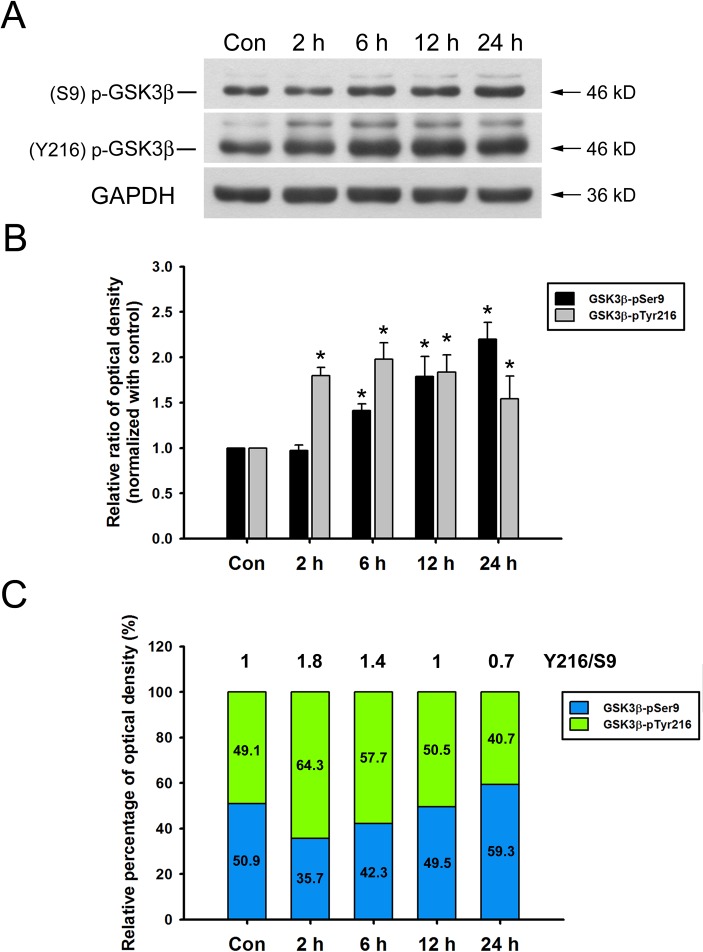
Fig 6. HYS-32 modulates GSK3β phosphorylation.
(A) Cell lysates from control astrocytes (Con) or astrocytes treated for 2, 6, 12, or 24 h with 5 μM HYS-32 were subjected to 10% SDS-PAGE, and analyzed by immunoblotting with antibodies against GSK3β-pTyr216, GSK3β-pSer9, or GAPDH. (B) Densitometric analyses of GSK3β-pTyr216 and GSK3β-pSer9 expressed as the density of the bands in the treated groups relative to the control. *p<0.05 compared to control using one-way ANOVA with Dunnett’s post-hoc test. The results were collected from five independent experiments. (C) According to the data in (B), the stacked bar graph showing the relative percentage of phospho-GSK3β at indicated time.

