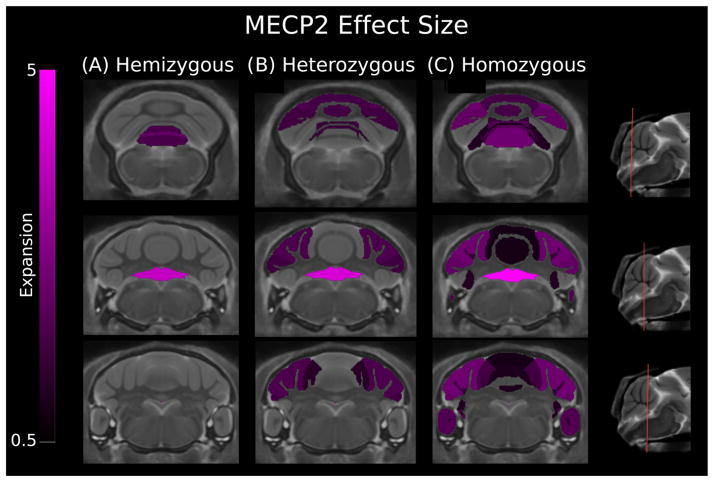
Figure 3.
Three coronal slices of the Methyl-CpG binding protein-2 (MECP2) mouse models compared with wild type showing effect size for cerebellum structures. The hemizygous model showed increases in the gray and white matter of vermis lobule IX and gray matter of vermis lobule X. The heterozygous model showed expansion of vermis lobule VI, VIII, and X. In the hemispheres, the anterior, simple, crus I, and crus II gray matter are larger in volume. The homozygous model had expansion of vermis lobules III–X, the anterior lobule, the paraflocculus, and simple, crus I, and crus II hemisphere lobules. A positive effect size represents an expansion of the region in the MECP2 mouse relative to controls (volumes have been normalized to total brain volume). These changes have been corrected for multiple comparisons using a false discovery rate threshold of 5%. Genotypes: hemizygous: −/y, heterozygous: −/+, homozygous: −/−.

