
Figure 2.
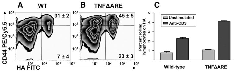
Enhanced HA binding and rolling flux fraction of CD4+ in TNFΔARE mice. (A and B) CD4+ T cells from the MLN of TNFΔARE mice and WT littermates were incubated with fluorescein isothiocyanate (FITC)-labeled HA (HA-FITC) and analyzed by flow cytometry. Unconjugated soluble HA was used as specificity control (data not shown). Cells were gated on forward scatter, side scatter, and CD4. Representative data are from 4 mice at 20 weeks of age. (C) Physiologic flow conditions were produced using a flow chamber at 2 dyn/cm2. Rolling interactions were analyzed using 107 cells in 8 or more fields of view (pooled mean ± SEM rolling fraction from 3 separate experiments; P < .05).