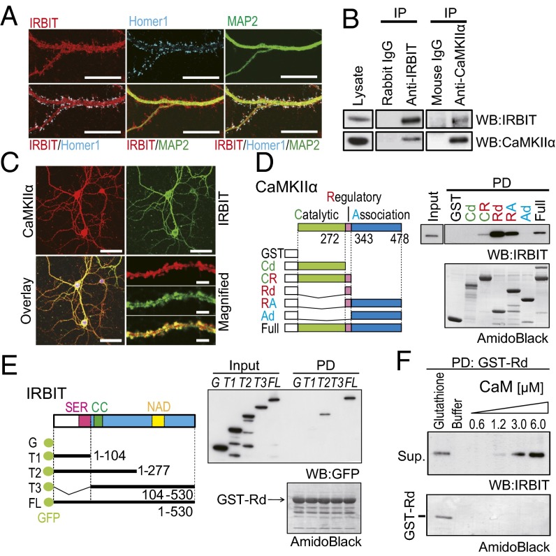
Fig. 1.
IRBIT interacted with CaMKIIα. (A) Cultured hippocampal neurons were stained with antibodies against IRBIT (red), Homer 1 (blue), and MAP2 (green). (Scale bar, 20 μm.) (B) Co-IP of IRBIT and CaMKIIα from the hippocampus. (C) The expression of CaMKIIα (red) and IRBIT (green) antibodies. (Scale bar, 50 μm.) The magnified images of the dendrite are shown. (Scale bar, 5 μm.) (D) Identification of CaMKIIα binding region to IRBIT in vitro. (Left) Schematic diagram of GST–CaMKIIα truncated mutants. CR, catalytic domain plus regulatory domain; Rd, regulatory domain; RA, regulatory domain plus associated domain; Cd, catalytic domain; Ad, associated domain. (Right) Pull-down assay using various GST–CaMKIIα mutants and purified IRBIT (from Sf9 cells). (E) Identification of the binding region of IRBIT to CaMKIIα. (Left) Schematic diagram of GFP–IRBIT truncated mutants. (Right) Pull-down assay using GST–Rd and GFP–IRBIT truncation mutants. (F) Ca2+–CaM and IRBIT competitively bind to CaMKIIα. Purified IRBIT was pulled down with GST–Rd and eluted with various concentration of Ca2+–CaM. Elution with glutathione was used as a positive control.

