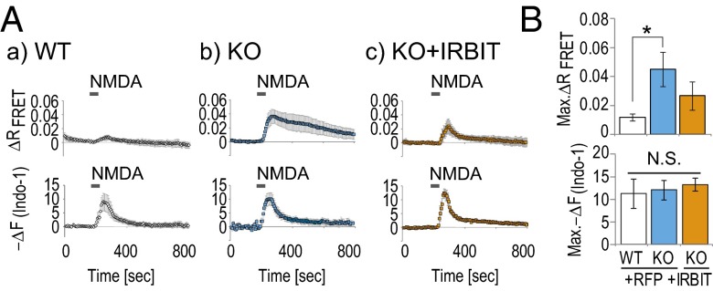
Fig. 4.
CaMKIIα activity is enhanced in hippocampal neurons from IRBIT KO mice. (A) Simultaneously imaging of FRET and Ca2+ change (Indo-1) after NMDA stimulation in WT or IRBIT KO cultured hippocampal neurons transfected with mRFP or IRBIT/mRFP. (B) Quantitation of FRET and Ca2+ peak amplitude [Max. −ΔF(Indo-1)] that were expressed as the averaged amplitude of 0–50 s are equal to zero.

