Fig. 6.
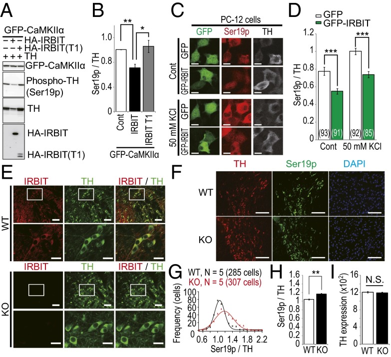
IRBIT regulates the phosphorylation level of tyrosine hydroxylase (TH) in the dopaminergic neurons of the VTA. (A) GFP–CaMKIIα cells were transfected with TH and HA–IRBIT or HA–IRBIT T1 fragment (1–104). After 24 h, cells were stimulated with 2.5 μM BrA for 2 min. (B) Quantitative analysis of phospho-TH in A. (C) PC-12 cells were transfected with GFP or GFP–IRBIT. After 24 h, cells were stimulated with 50 mM KCl for 10 min. Nonstimulated and KCl-stimulated cells were fixed and stained with indicated antibodies. (D) Quantitative analysis of phospho-TH in C. Total number of cells is indicated in each bar. (E) Adult WT and IRBIT KO mice brain sections were stained with anti-IRBIT (red) and anti-TH (green) antibodies. (Scale bar, 50 μm.) The boxed regions are shown at increased magnification. (Scale bar, 20 μm.) (F) Adult WT and IRBIT KO mice brain sections were stained with anti-TH (red), anti–phospho-TH (Ser19p, green) antibodies, and DAPI (blue). (Scale bar, 100 μm.) (G–I) Quantitative analysis of phospho-TH levels of dopaminergic neurons in the VTA of IRBIT KO mice. (G) Histogram of normalized TH phosphorylation. (H) Average of normalized TH phosphorylation. (I) Average of TH expression. n = 5. Total cell numbers of quantified TH positive neurons were 285 (WT) and 307 (KO).

