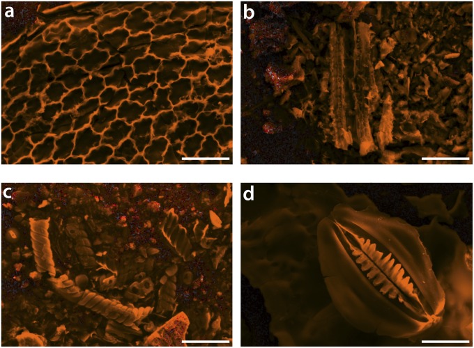
Fig. 3.
Secondary electron images of silica bodies (grayscale) overlaid with Si maps from energy-dispersive X-ray spectroscopy (orange). (Scale bars, 25 μm.) (A) Selaginella sp., (B) Gnetum gnemon, (C) Ephedra californicum, and (D) Equisetum hyemale. Some distinct mineralized plant tissues can be recognized: in A, epidermal cell walls are silicified; in C, possible silicified vascular tissue; and in D, a silicified stomatal complex. It is noteworthy that these tissues are all near the sites of transpiration.

