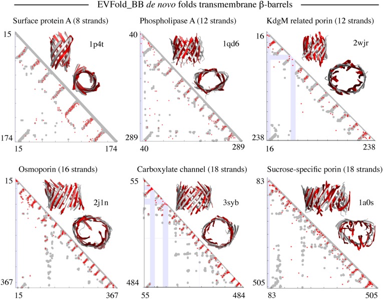
Fig. 3.
Blinded benchmark de novo 3D models of transmembrane β-barrels. Shown are predicted contact maps (red, ECs; gray, crystal contacts ≤ 5 Å; blue, gaps in crystal structure) and front and top views of folded structures (red, de novo folded; gray, crystal structure) for six proteins in the dataset.

