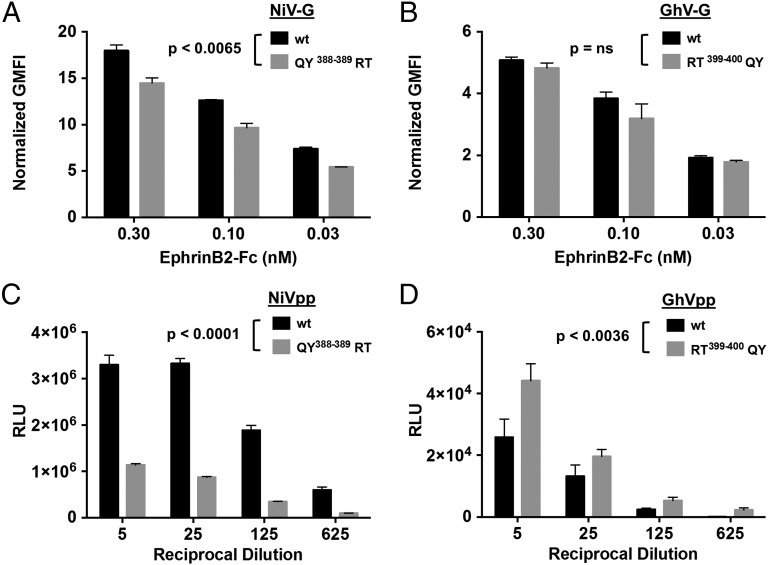
Fig. 5.
GhV-G uses ephrinB2 less efficiently. (A and B) EphrinB2 binding to WT NiV-G (A) or GhV-G (B) and the respective QY388-389 (A) or RT399-400 (B) mutants involving the secondary ephrinB2-binding interface. Soluble EphrinB2–Fc binding to cell surface HNV-G was background subtracted and normalized to HNV-G cell-surface expression as detected by anti-HA staining. Data are shown as the mean normalized geometric mean fluorescent intensity (GMFI) ± SD of duplicate experiments. One representative experiment of three is shown. (C and D) Effects of reciprocal NiV-G (QY388-389RT) and GhV-G (RT399-400QY) mutations in the context of an infectivity assay using VSV-based pseudotyped particles, prepared as described in Materials and Methods. (C and D) NiVpp (C) and GhVpp (D) bearing WT F/G or the indicated mutant G proteins were used to infect U87 glioblastoma target cells over a range of viral inoculum (indicated by a dilution series of the viral stock preparation). Entry of NiVpp/GhVpp results in production of luciferase by the reporter virus backbone (VSV-ΔGLuc). Data are shown as mean ± SD from quadruplicate samples. One representative experiment of two is shown. QY388-389RT mutations in NiV-G (C) and RT399-400QY mutations in GhV-G (D) significantly decreased (P < 0.0001) and increased (P < 0.0036) infectivity with respect to their parental WT NiVpp and GhVpp (two-way ANOVA for multiple comparisons across the dilution series examined). In contrast, only the NiV-G QY388-389RT mutant (A) showed moderate but significantly decreased ephrinB2 binding with respect to WT NiV-G (P < 0.0065; two-way ANOVA for multiple comparisons). Expression of the various F and G proteins on pseudotyped particles and the cell surface are shown in SI Appendix, Fig. S7.

