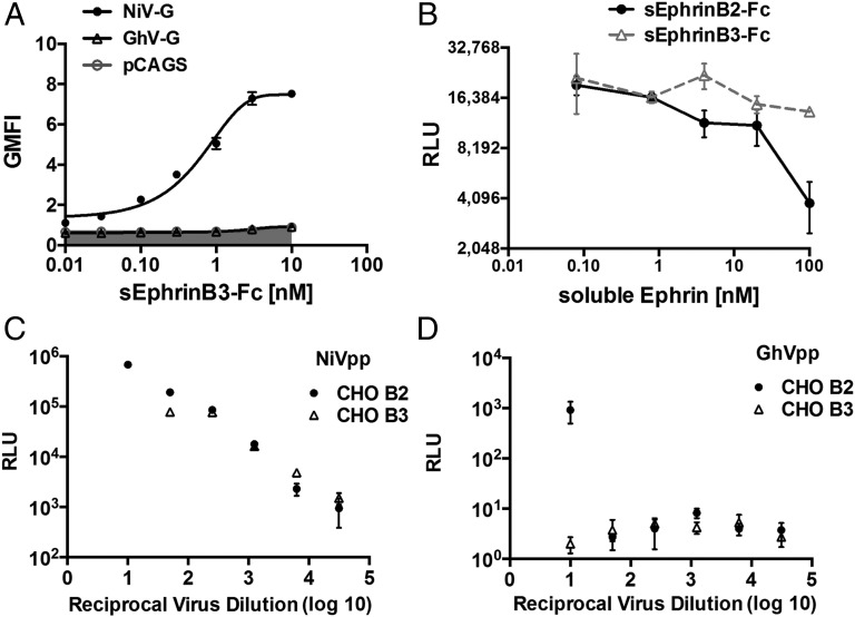
Fig. 6.
GhV-G does not use ephrinB3 as a cell-surface receptor. (A) Soluble ephrinB3–Fc binds NiV-G but not GhV-G. NiV- or GhV-G was transfected into CHO cells. At 18 h after transfection, cells were stained with increasing amounts of recombinant soluble ephrinB3–Fc and processed for FACS analysis after secondary staining with PE-conjugated anti-human Fc antibodies. Data are shown as mean ± SD of experiments performed in duplicates. One representative experiment of two is shown. (B) sEphrinB2–Fc but not sEphrinB3–Fc, blocks GhVpp infection of U87 cells. GhVpp prepared as described in Fig. 5 was used to infect U87 cells in the presence of increasing amounts of soluble ephrinB2–Fc or ephrinB3–Fc as indicated. Data are shown as mean ± SD of quadruplicate infection experiments. (C and D) NiVpp (C) and GhVpp (D) were used to infect CHO cells stably expressing ephrinB2 (CHO-B2) or ephrinB3 (CHO-B3) over a fivefold dilution series of viral inoculum ranging from 1:10 to 1:31,250 (reciprocal virus dilution indicated on the x axis). Entry of NiVpp/GhVpp results in production of luciferase by the reporter virus backbone (VSV-ΔGLuc). Data are shown as mean ± SD from quadruplicate samples. One representative experiment of two is shown.

