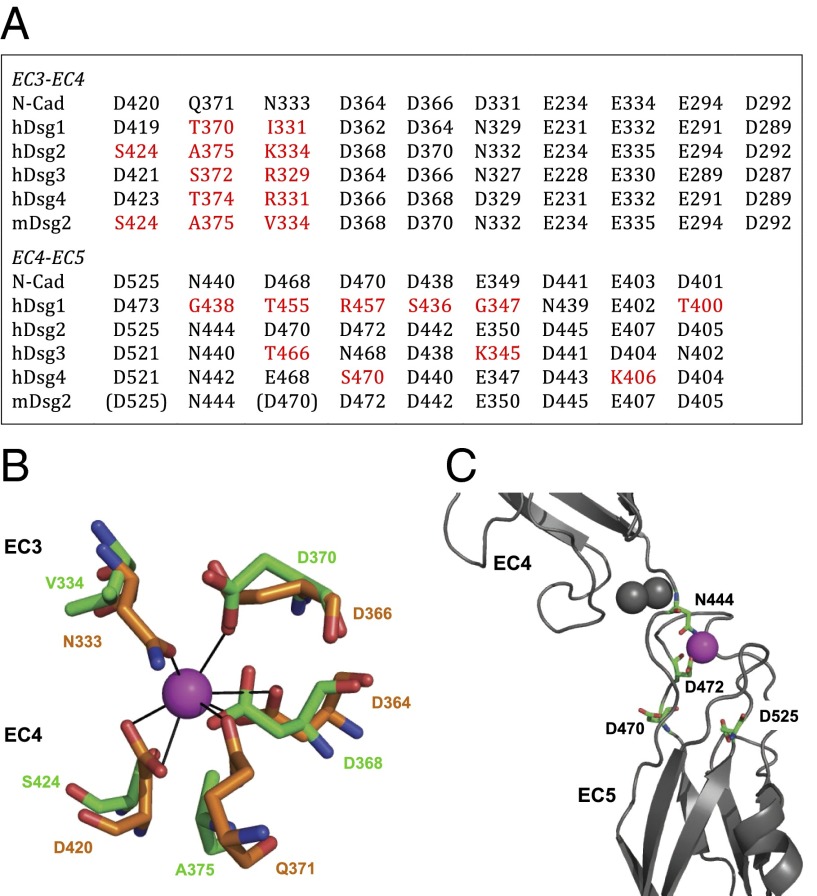
Fig. 4.
Calcium coordination in Dsg2 is not conserved. (A) Table listing the residues involved in EC3–EC4 and EC4–EC5 interdomain calcium coordination. Nonconservative substitutions in mDsg2 and hDsgs are in red; residues located too far for proper coordination are in brackets. (B) Side chain coordination of one of the three Ca2+ ions at the EC3–EC4 region; residues coordinating the same Ca2+ ion (purple) in N-cadherin (PDB ID code 3Q2W, orange chain) and in the mDsg2 homology model in green. (C) Coordination of one of the three Ca2+ ions at the EC4–EC5 region; the two Ca2+ binding residues D470 and D525 located in loop regions are too distant from the calcium ion, leaving only residues N444 and D472 available for coordination. Gray spheres are the other two Ca2+ ions.

