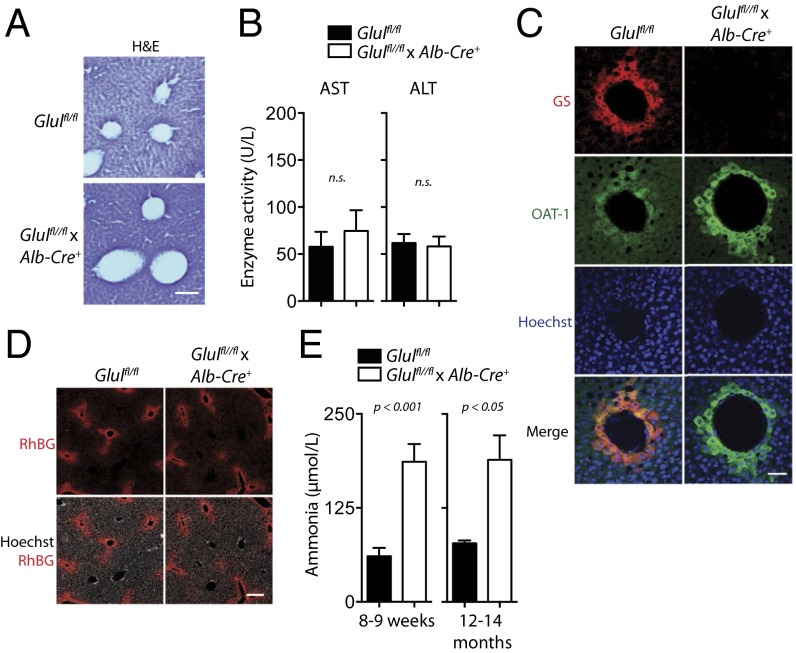
Fig. 2.
Intact liver architecture/zonation and elevated systemic ammonia levels by liver-specific deletion of GS. (A) Representative H&E-stained sections of snap-frozen liver tissue obtained from Glulfl/fl × Alb-Cre+ and Glulfl/fl mice of n = 3 is shown. (Scale bar: 100 μm.) (B) Asp aminotransferase (AST) and Ala aminotransferase (ALT) activity was determined in the serum of Glulfl/fl (n = 10) and Glulfl/fl × Alb-Cre+ mice (n = 12). n.s., not statistically significantly different. (C) Immunofluorescence analyses of snap-frozen liver tissue from Glulfl/fl × Alb-Cre+ (Lower) and Glulfl/fl (Upper) mice were performed for GS (red), ornithine aminotransferase (OAT; green), and Hoechst 34580 (blue). One representative set of images of n = 3 is shown. (Scale bar: 20 μm.) (D) Immunofluorescence analyses of snap-frozen liver tissue from Glulfl/fl × Alb-Cre+ (Right) and Glulfl/fl (Left) mice were performed for ammonia transporter Rh family B glycoprotein (RhBG; red) and Hoechst 34580 (gray). One representative set of images of n = 3 (Glulfl/fl) and n = 4 (Glulfl/fl × Alb-Cre+) is shown. (Scale bar: 200 μm.) (E) Ammonia levels were assessed in blood samples collected by cardiac puncture from 8- to 9-wk-old Glulfl/fl × Alb-Cre+ mice and Glulfl/fl mice (Left, n = 7–8, respectively) and from 12- to 14-month-old animals (Right, n = 3).

