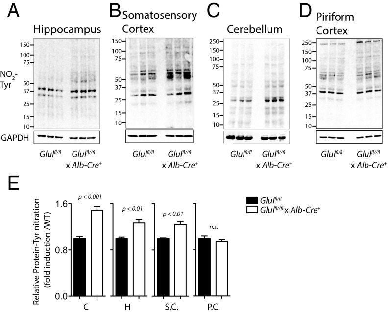
Fig. 3.
Hepatic GS KO triggers PTN in mouse brain. Protein samples harvested from brain slices of the cerebellum, hippocampus, somatosensory cortex, and piriform cortex of Glulfl/fl and Glulfl/fl × Alb-Cre+ mice were tested for PTN using Western blot analysis. (A–D, Upper) Representative blots showing anti–3′-nitrotyrosine immunoreactivity. (A–D, Lower) GAPDH served as a loading control. (E) Mean ± SEM of the densitometry of the cerebellum (C), hippocampus (H), somatosensory cortex (S.C.), and cortex piriform (C.P.) is presented (n = 6 for Glulfl/fl and n = 9 for Glulfl/fl × Alb-Cre+ mice for the cerebellum and hippocampus, n = 6 for Glulfl/fl and n = 8 for Glulfl/fl × Alb-Cre+ mice for the somatosensory cortex, and n = 3 for Glulfl/fl and n = 6 for Glulfl/fl × Alb-Cre+ mice for the cortex piriform).

