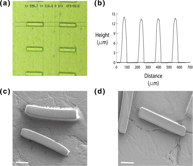
Fig. 2.

Microrod dimension and structure. a A phase microscopy shows microrods with approximately 15 μm width and 100 μm length; b The microstructure height was measured approximately 15 μm using an Ambios Technology XP-2 profilometer (note: the x axis distance value in this figure does not represent the actual width of the microrods); SEM images represent the structure of empty microrods c and MGF microrods d. Scale bar, 20 μm
