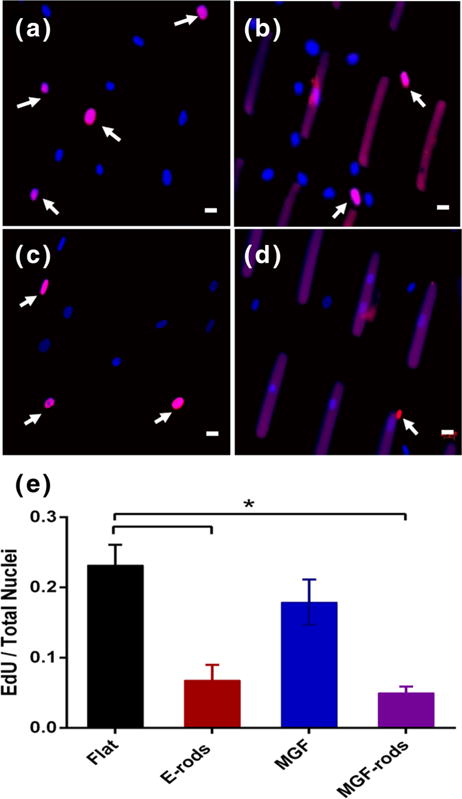
Fig. 5.

Microrods blunt proliferation of hMSCs. Newly dividing cells (pink) vs. non-dividing cells (blue) on (a) flat surfaces, (b) with empty microrods (E-rods), (c) on flat surface with MGF in the media, or (d) with MGF eluting microrods (MGF-rods), as seen by fluorescence microscopy. (e) EdU/total nuclei per condition show that microrods inhibit new synthesis of DNAwith or without MGF. Dividing nuclei (arrows) stained with EdU (pink), non-dividing with DAPI (blue), and microrods artificially stained with DAPI and EdU (Purple). Mean ± SE, n=4, * p<0.05. Scale bar, 20 μm
