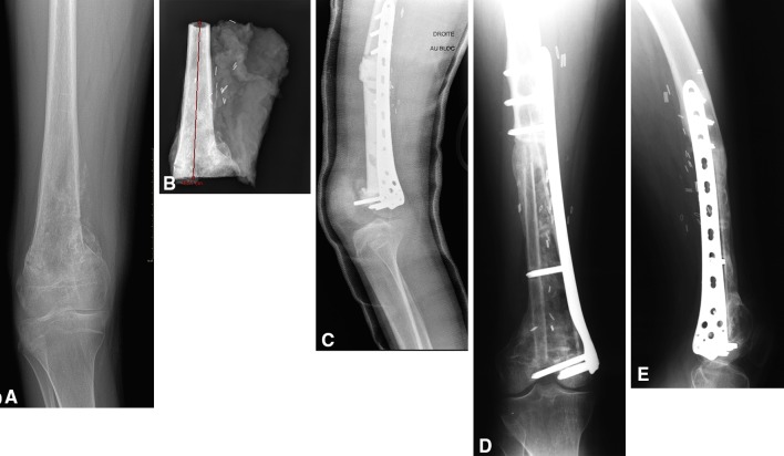
Fig. 2A–E.
(A) An AP radiographic view shows a right distal femur osteosarcoma (Patient 7). (B) The distal femur resection with epiphyseal preservation can be seen on this radiograph. The red line represents the measurement of the bony resection, which in this case was 16.11 cm. (C) A postoperative lateral radiographic view shows the cement spacer in the bed of the resection stabilized with a locking plate.) (D) AP and (E) lateral radiographic views obtained 2 years after the second stage surgery show the reconstructed femur with the locking plate on the medial side and the nonvascularized fibula on the lateral side of the reconstruction. Knee flexion was 90°.

