Fig. 4.

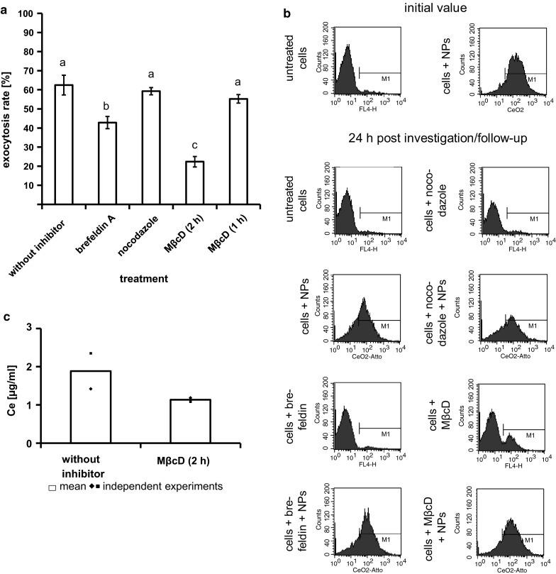
Strong inhibitory effect of MβcD and brefeldin A indicates the important role of plasma membrane cholesterol and Golgi-to-cell-surface-transport, respectively, during nanoparticles exocytosis. a The average exocytosis rate of CeO2 nanoparticles (treatment dose 1 µg/ml for 24 h) within 24 h was 62 ± 5 %. Nocodazole led to no obvious inhibition of nanoparticle exocytosis (exocytosis rate: 59 ± 2 %). The highest inhibition of exocytosis was caused by MβcD with an exposure time of 2 h indicating an important role of plasma membrane cholesterol for exocytosis. Brefeldin A treatment resulted also in an inhibition of exocytosis revealing an involvement of Golgi-to-cell-surface-transport in exocytosis process. Different letters indicate significant differences (P ≤ 0.05) between the various treatments. n ≥ 3 independent experiments; b Histograms of a representative flow cytometry analysis; NPs: nanoparticles. c The determination of cerium (Ce) in the supernatant of HMEC-1, which were exposed to nanoparticles for 24 h, revealed 24 h after washing and cell culture medium exchange (nanoparticle free medium) a higher amount of Ce than the supernatants of HMEC-1 which were additionally treated with MβcD-containing cell culture medium after washing and medium exchange. This confirmed the inhibition of exocytosis by MβcD. The Ce content in supernatants of cells, which were not treated with nanoparticles, was below the detection limit. n = 2 independent experiments
