
Fig. 2.
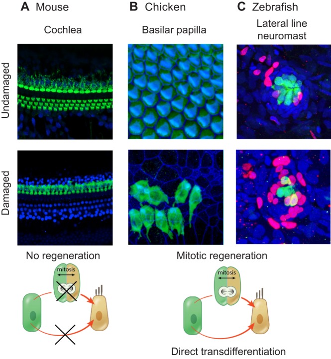
Regenerative capacities of the adult mouse cochlea, the chicken basilar papilla and the zebrafish lateral line system. (A) No sensory hair cell regeneration occurs in the adult mouse cochlea. Shown are the basal turns of postnatal day (P) 28-30 cochleae from control (undamaged) mice and from mice that were treated with sisomicin and furosemide at P21. Immunostaining was performed to detect myosin VIIa (green) and Sox2 (blue); the reduction in myosin VIIa-labeled hair cells indicates that the damaged hair cells are not able to regenerate. (B) Hair cell regeneration in the chicken basilar papilla. Organs were harvested from control chicks and from those that had received daily injections of streptomycin in the previous week. Myosin VIIa-labeled hair cells are in green and phalloidin staining (F-actin) is in blue. Image courtesy of M. Warchol. (C) Hair cell regeneration in the zebrafish lateral line system. Images show posterior lateral line neuromasts from control zebrafish and from those that were damaged with neomycin. Hair cells are labeled by GFP in green, while BrdU (red) marks proliferating cells and DAPI staining (blue) marks nuclei. Regeneration in the neuromast populations, as indicated by the number of BrdU-positive cells, occurs primarily through mitotic regeneration. Image courtesy of T. Piotrowski and A. Romero-Carvajal.