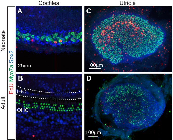
Fig. 3.

Comparison of the modes and capacity of hair cell regeneration in the neonatal and adult mouse cochlea and utricle. (A) Limited mitotic hair cell regeneration occurs in the neonatal cochlea following diphtheria toxin-induced damage. Shown is the apical turn of the cochlea from P7 Pou4f3DTR/+ mice, which received diphtheria toxin at P1 to ablate hair cells. Immunostaining for myosin VIIa (green) highlights hair cells, while Sox2-positive supporting cells are in blue. The presence of hair cells labeled with EdU (red) indicates mitotic hair cell regeneration. Image courtesy of R. Chai. (B) Conversely, no mitotic response (EdU labeling) or hair cell regeneration occurs in the damaged adult cochlea. The image shows a cochlea from Pou4f3DTR/+ mice that received diphtheria toxin one week previously. There is a complete loss of inner hair cells (IHC) and a partial loss of outer hair cells (OHC; green). (C) By contrast, robust mitotic hair cell regeneration (as indicated by the abundance of EdU-labeled hair cells) is observed in the neonatal utricle. The image is of a utricle from P30 Pou4f3DTR/+ mice that received diphtheria toxin at P1 to ablate hair cells. Image courtesy of R. Chai and T. Wang. (D) Scant proliferation and regeneration are observed in the damaged mature utricle. Shown is a utricle from an adult mouse previously treated with a vestibulotoxin.
