Fig. 3.
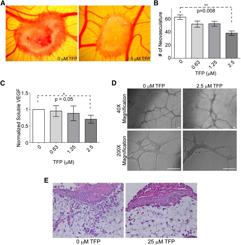
TFP reduces angiogenesis and invasion examined by a chicken CAM assay. (A) Representative image of a CAM assay demonstrates that treatment of HT1080 cells with TFP reduces the ability of those cells to induce angiogenesis. (B) Quantification of neovasculature induced by HT1080 cells in the CAM assay; 2.5 μM treatment results in a significant decrease in angiogenesis. (C) TFP treatment reduces soluble VEGF. HT1080 cells treated with various doses of TFP demonstrate a reduction in VEGF expression as determined by VEGF ELISA. (D) TFP treatment does not induce changes in HUVEC network formation on a three-dimensional Matrigel platform. HUVEC cells treated with 2.5 μM TFP do not have impaired network formation or change in morphology compared with the vehicle-treated endothelial cells. Bar = 100 μm. (E) Hematoxylin and eosin staining of a tissue section collected from the CAM assay shows that HT1080 cells implanted on the CAM surface can invade through the underlying membrane, whereas TFP treatment reduces the ability of these cells to invade but continue to grow.

