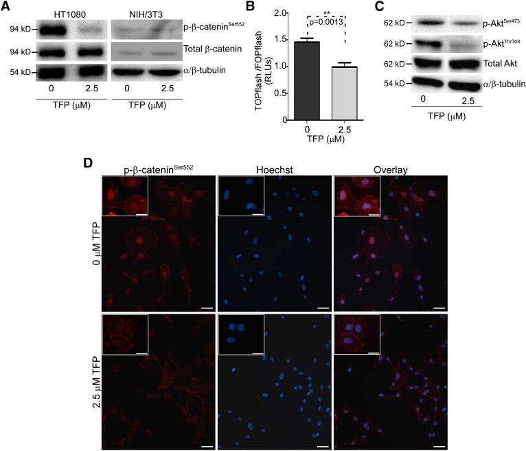
Fig. 4.
TFP treatment of HT1080 cells results in a decrease in phosphorylated β-cateninSer552, AKTSer473, and AKTThr308. (A) Western blot analysis of HT1080 lysates shows that TFP treatment results in a decrease in p-β-cateninSer552, whereas there are no changes in TFP-treated NIH/3T3 cells. Total β-catenin and α/β tubulin were used as controls. (B) TFP treatment of HT1080 cells reduces the transcriptional activity of β-catenin as assessed by TOPflash/FOPflash luciferase activity, expressed in relative luciferase units (RLUs). (C) Decreases of phosphorylated AKT in HT1080 cells treated with TFP (2.5 μM) for 18 hours examined by Western blot using antiphospho-AKTSer473 and AKTThr308 antibodies, respectively. Total AKT and α/β-tubulin were used as controls. (D) Immunofluorescent staining of HT1080 cells treated with TFP shows decreased nuclear p-β-cateninSer552 staining compared with vehicle control using anti–phospho-β-catenin antibody. Nuclei were stained by Hoechst. β-Catenin staining and nuclear staining were superimposed (overlay) Bar = 50 μm. Enlarged representative images are shown in the inserts. Bar = 20 μm.

