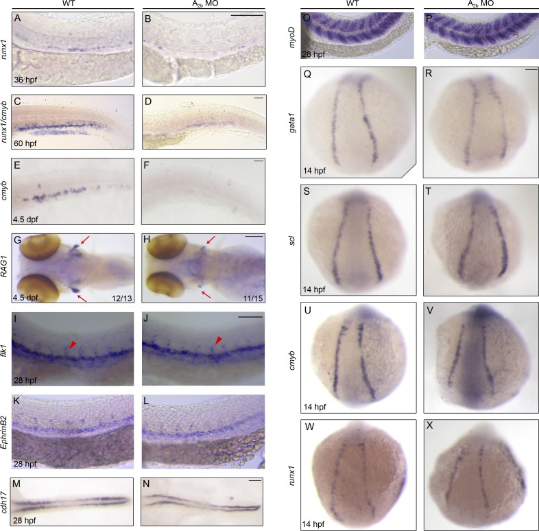
Figure 3.
Adenosine signaling through A2b specifically regulates HSPC development. (A–H) Control or A2b MO–injected embryos stained for runx1 in the AGM at 36 hpf (A and B), runx1/cmyb in the CHT at 60 hpf (C and D), cmyb in the CHT (E and F), and RAG1 in the thymus (red arrows in G and H) at 4.5 dpf. (I–X) Control or A2b MO–injected embryos stained for vasculature (flk1; I and J), dorsal aorta (ephrinB2; K and L), pronephros (cdh17; M and N), and somite (myoD; O and P), primitive hematopoiesis (gata1 and scl; Q–T), and cmyb and runx1 (U–X) at the developmental stages indicated. In I and J, red arrowheads mark intersegment staining. Bars, 100 µm.

