Figure 6.
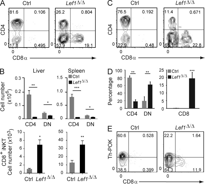
LEF1 prevented CD8α expression on peripheral iNKT cells. (A) CD4 and CD8 expression on Tetr+TCRβ+ iNKT cells from the liver of control or Lef1Δ/Δ mice was determined by flow cytometry. (B) The mean number of CD4+, DN, and CD8+ iNKT cells from the indicated tissues in control and Lef1Δ/Δ mice. Data were averaged from at least six independent experiments. (C) CD45.1+CD45.2+ control and CD45.1−CD45.2+ Lef1Δ/Δ FACS-sorted LK bone marrow cells were mixed at 1:1 ratio and transferred into lethally irradiated CD45.1+ C57BL/6 mice. 6–8 wk later, reconstituted mice were analyzed by flow cytometry for CD4 and CD8 expression on control or Lef1Δ/Δ iNKT cells from the liver. (D) The mean percentage of the CD4+, DN, and CD8+ liver iNKT cells from competitive BM chimeras. Data are representative of three independent experiments with two to three chimeras each. (E) Th-POK and CD8α expression in splenic iNKT cells from the indicated mice, as determined by flow cytometry. Data are representative of five independent experiments with one to two mice per genotype. All error bars represent SEM. *, P < 0.05; **, P < 0.01; ***, P < 0.001.

