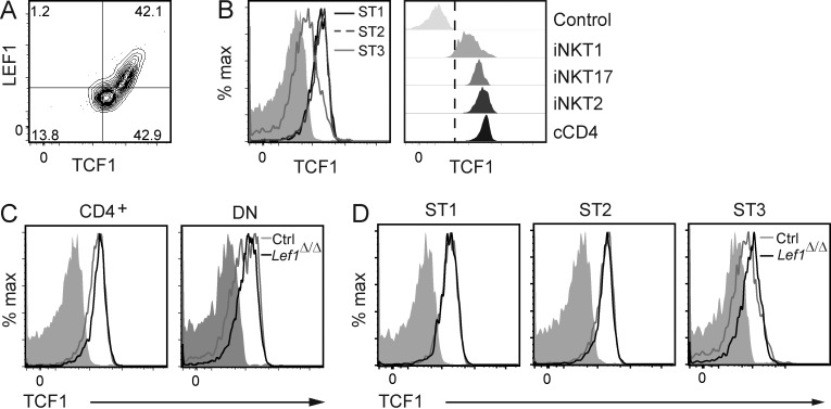
Figure 7.
TCF1 expression was not affected by deletion of Lef1 in iNKT cells. (A) Expression of LEF1 and TCF1 in total WT FVB/NJ iNKT cells, as determined by flow cytometry. (B) TCF1 expression in the indicated iNKT stages (left) or iNKT subsets (right) from WT FVB/NJ thymocytes was determined by flow cytometry and is shown as histograms. Solid gray or control represents anti-TCF1 staining in TCF1-deficient iNKT cells and serves as a negative control. (C and D) TCF1 expression in CD4+ and DN (C) or ST1, ST2, and ST3 (D) thymic iNKT cells from control and Lef1Δ/Δ mice was determined by flow cytometry. Data are representative of four independent experiments.

