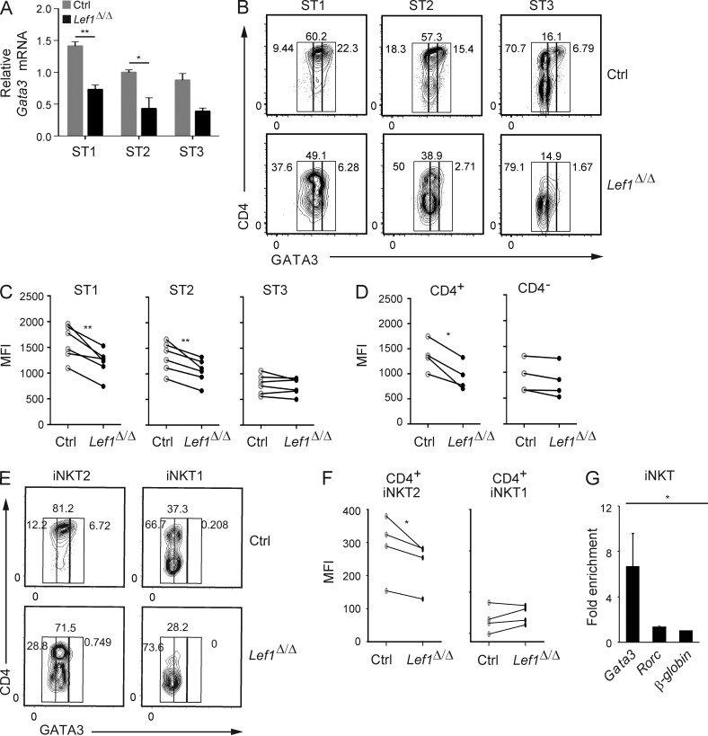
Figure 8.
LEF1 was required for proper expression of GATA3 in CD4+ iNKT cells. (A) Expression of Gata3 mRNA relative to Hprt in sorted ST1, ST2, and ST3 iNKT cells from control and Lef1Δ/Δ mice was determined by qRT-PCR. Data were combined from three independent experiments. (B) GATA3 versus CD4 expression in ST1, ST2, and ST3 thymic iNKT cells from control and Lef1Δ/Δ mice, as determined by flow cytometry. (C and D) MFI for GATA3 in ST1, ST2, and ST3 iNKT cells (C) or in CD4+ and CD4− iNKT cells (D) from the thymus of control and Lef1Δ/Δ mice. (E) GATA3 versus CD4 expression in iNKT1 (PLZFloTBET+) and iNKT2 (PLZFhiTBET−) cells from control and Lef1Δ/Δ mice, as determined by flow cytometry. Data are representative of four independent experiments. (F) MFI for GATA3 in thymic CD4+ iNKT2 and CD4+ iNKT1 cells from the indicated mouse strains. (C, D, and F) Each circle represents one mouse (paired Student’s t test: *, P < 0.05; **, P < 0.01). (G) LEF1 chromatin binding was determined by a-LEF1 ChIP on sorted thymic ST1/ST2 Vα14Tg iNKT cells, followed by qPCR using primers spanning a TCF1/LEF1-binding site upstream of the Gata3b promoter, the Rorc promoter, or an unrelated sequence at the β-globin gene locus. Numbers indicate mean fold enrichment of the immunoprecipitated DNA relative to the β-globin locus from three independent experiments is shown. Error bars represent SEM. *, P < 0.05; **, P < 0.01.

