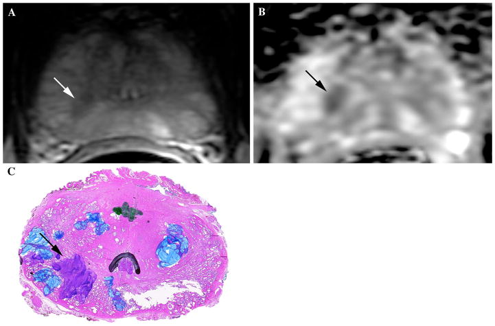
Fig. 2.
61-year-old male with elevated PSA value (6.1 ng/mL at time of mpMRI exam) and prior TRUS-guided biopsy showing moderate-high volume Gleason 5+4 (9) tumor on the right in 4/6 cores and low volume Gleason 3+3 (6) tumor on the left in 2/6 cores. Patient was referred for mpMRI with endorectal coil for pre-operative planning. A Axial T2W image at the level of the midgland shows an area of decreased T2 signal intensity in posterior medial right PZ (white arrow). B Axial ADC map shows corresponding area of markedly restricted diffusion and low ADC value (black arrow). This lesion was considered to be the index lesion and felt to represent a Gleason 7+ tumor in light of the markedly restricted diffusion. C Whole mount histology image at this location within the prostate confirms the presence of an index lesion (black arrow) with components of Gleason 4 (purple) disease, corresponding with findings on initial biopsy and mpMRI. Of note, the blue marking on the image reflects prostatic atrophy and the green marking in the center refers to a small low-grade focus of prostate cancer that was not deemed to be the index cancer. This case is an example of one used in the dedicated reader education program.

