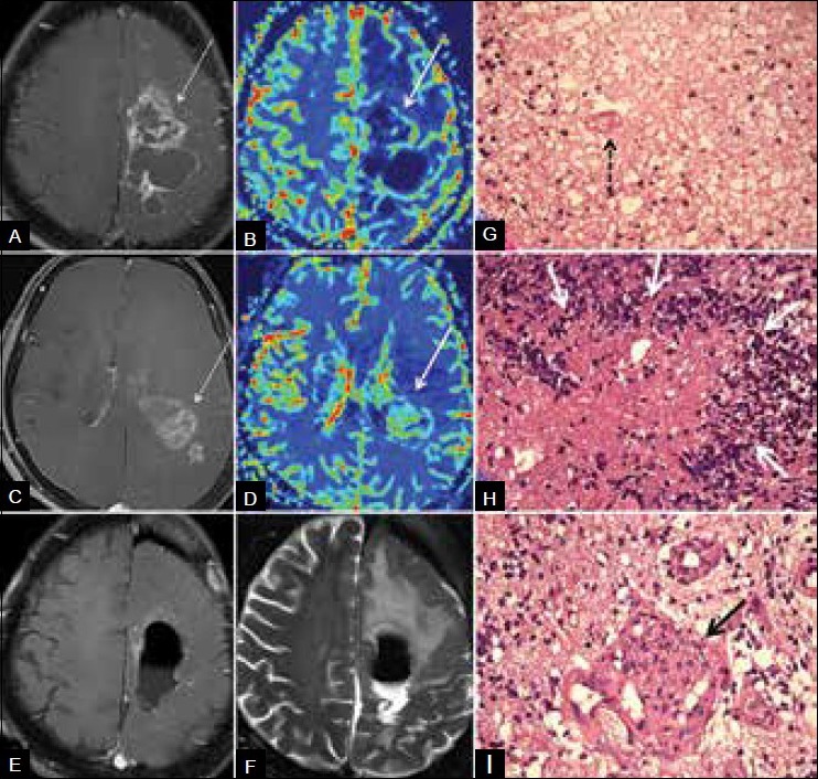
Figure 2B (A-I).

Mixed radiation necrosis and recurrence. Three years after surgery and RT–CT for grade 2 astrocytoma, patient presented with new multifocal mass. (A) Contrast (B) perfusion images show hypoperfused enhancing lesion (arrow) near the tumor bed, suggestive of radiation necrosis. (C and D) Caudal section in the same patient shows hyperperfused enhancing (arrows) tumor recurrence. (E and F) Postoperative MRI shows excision. (G) Histology from the tumor bed mass (H and E, ×40) shows RT necrosis comprising fibrinoid material with extravasated RBCs, inflammatory cells, and fibrinoid vasculopathy (arrow). No palisading of the necrosis is seen. (H) Histology from caudal lesion (H and E, ×40) shows recurrence with palisading of tumor necrosis by tumor cells forming wreath rosettes (arrows). (I) Histology from caudal lesion (H and E, ×40) shows recurrence with increased vascularity and endothelial proliferation giving glomeruloid appearance (arrow)
