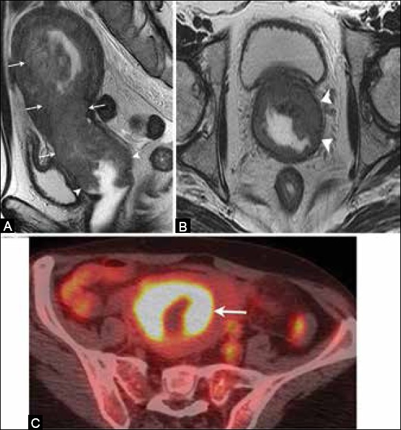
Figure 14 (A-C).

A 62-year-old female with endometrial cancer. (A) Sagittal and (B) axial T2W MR images show an endometrial mass (arrow) extending inferiorly and involving the vagina (arrowheads) indicating stage IIIB disease. (C) FDG-PET/CT demonstrates increased FDG uptake by the tumor (arrow)
