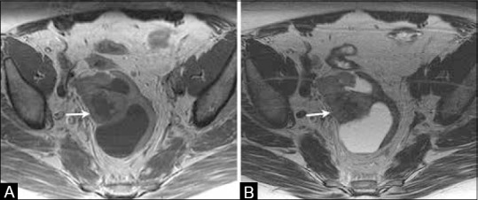
Figure 19 (A and B).

A 69-year-old female with history of treated endometrial cancer surgically treated with hysterectomy. (A) Axial T1W and (B) T2W MR images show a complex cystic solid lesion in the pelvic region suggestive of recurrent disease

A 69-year-old female with history of treated endometrial cancer surgically treated with hysterectomy. (A) Axial T1W and (B) T2W MR images show a complex cystic solid lesion in the pelvic region suggestive of recurrent disease