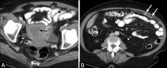
Figure 4 (A and B).

A 75-year-old female with endometrial cancer. (A) Axial contrast-enhanced computed tomography image of the pelvis shows the thick hypodense endometrium (black arrow). (B) Axial contrast-enhanced CT image shows peritoneal implants (white arrows) in this patient with clear cell endometrial carcinoma
