
Figure 7 (A and B).
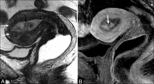
A 68-year-old female with endometrial cancer. (A) Sagittal T2W image showing a hypointense mass (black arrow) distending the endometrial cavity with integrity of the junctional zone (white arrows). (B) T1W post-contrast image shows heterogeneous enhancement of the endometrial cavity (arrow) with no evidence of myometrial invasion or cervical involvement indicating stage IA disease