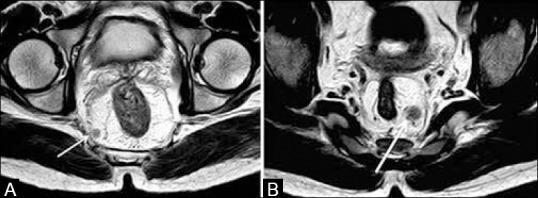
Figure 15 (A and B).

Axial T2W MRI. (a) Tumor deposit in the right mesorectal fat (arrow) located <1mm from the MRF (CRM +ve). (b) Left mesorectal node (arrow), 1-2 mm from the MRF indicating a threatened CRM. Node has heterogeneity and irregular margins
