Figure 20 (A and B).

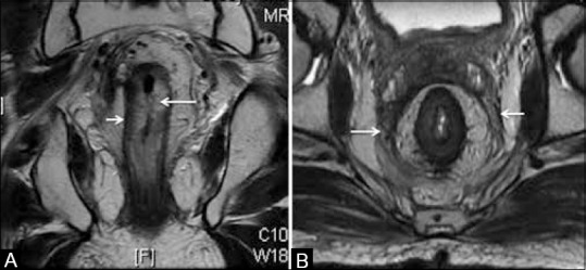
Post RT appearances. (A) Coronal T2W MRI shows thickened hyperintense submucosa due to edema (long arrow) with the intact muscularis (short arrow). (B) Axial T2W MRI with diffusely thickened mesorectal fascia (arrows)

Post RT appearances. (A) Coronal T2W MRI shows thickened hyperintense submucosa due to edema (long arrow) with the intact muscularis (short arrow). (B) Axial T2W MRI with diffusely thickened mesorectal fascia (arrows)